A team of clinician researchers from Lawson Health Research Institute have partnered with medical device company CoapTech LLC to study a new method of feeding tube insertion. The study is assessing the safety and efficacy of a new device called the PUMA-G System with 25 patients at London Health Sciences Centre (LHSC). The new method may offer improved patient safety while providing cost savings to the health care system.
In October 2018, the team was the first in the world to use the device when they inserted a feeding tube for Sonny McGlone. Sonny, a 76-year-old man from Sarnia, Ontario, was being treated with radiation therapy for head and neck cancer.
“I had seven weeks of radiation which killed my taste buds. I couldn’t swallow or eat and I was rapidly losing weight,” explains Sonny. “I was pleasantly surprised by the feeding tube procedure. While the tube was obviously inconvenient, it was a life saver.”
Feeding tubes, also called gastrostomy tubes, are needed by those who cannot maintain adequate nutrition through the mouth. They can be very important to the treatment and recovery of increasingly common conditions such as cancer, stroke and trauma.
The insertion of a feeding tube is conventionally guided by x-ray imaging or endoscopy, a procedure that uses a camera and light to visualize the stomach. While highly effective, these methods require use of specialized imaging suites that are critically needed by many patients. The time and resources required in these suites can also be costly to the health care system.
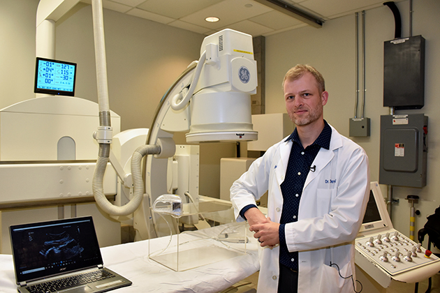
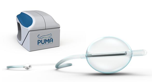
The PUMA-G System provides a new method of feeding tube insertion that can be performed at a patient’s bedside. The device uses a magnetic balloon that is fed through a patient’s mouth and guided to the stomach with an external magnet. The balloon is inflated with water and ultrasound guides the treating physician as they insert a needle through the stomach and into the balloon. The balloon catches a wire that is then pulled back up and out the mouth as the balloon is removed. A feeding tube can then be pushed back down over the wire and safely out the stomach.
“This new method is already showing promise as being safe, effective and efficient. It allows feeding tubes to be inserted at the patient’s bedside and reduces the demand on specialized imaging suites,” says Dr. Derek Cool, Associate Scientist at Lawson and Interventional Radiologist at LHSC. “This could be especially important for patients in the intensive care unit (ICU). They can benefit from the safety of that environment without being moved.”
The PUMA-G System also minimizes the risk of puncturing other organs. Traditional methods do not show the space between the skin and stomach. While rare, there’s a small risk that organs like the liver or colon could be injured during insertion. Ultrasound minimizes this risk by allowing physicians to see between the skin and stomach while the needle is being inserted.
The device has potential to provide visualization for procedures in other hollow organs too.
“The PUMA-G System is only our first application of a using ultrasound and magnets to guide procedures in hollow organs,” says Howard Carolan, CEO of CoapTech. “This study aims to demonstrate the overall benefits of switching to what we believe will be the new standard of care for feeding tube procedures.”