3D imaging technology could improve outcomes for patients with breast cancer
A study at Lawson is looking to determine if digital breast tomosynthesis, a type of 3D imaging, is better at detecting breast tissue abnormalities than the 2D mammography regularly used today.
During a conventional digital 2D mammogram, two x-ray images are taken of the breast, one from top-to-bottom and another from side-to-side at an angle. This technology is limited by the overlapping breast tissue that occurs from the required compression of the breast, and breast abnormalities may be hidden.
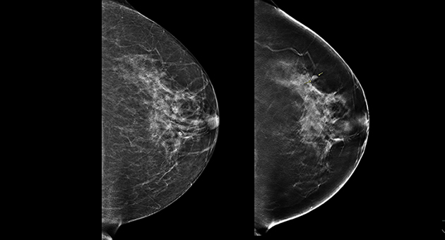
(Left) 2D mammogram image of left breast, where no lesion was visible. (Right) 3D tomographic image of the same breast showing a lesion, indicated by arrows.
A tomosynthesis exam is relatively new technology in which the x-ray tube moves in an arc over the compressed breast and captures multiple images from different angles. The images are then reconstructed into a set of 3D images by a computer. By being able to examine the breast at multiple layers of depth, the radiologist is better able to distinguish normal breast tissue from potential abnormalities. It is therefore assumed that tomosynthesis may solve some challenges associated with standard mammography, and could be especially useful for women with dense breast tissue.
In the Tomosynthesis Mammographic Imaging Screening Trial (TMIST), women are randomized to receive screening with standard digital 2D mammography, or digital 2D mammography plus tomosynthesis. Participants will undergo either an annual or biennial screening frequency, depending on their risk factors for breast cancer, for approximately four years. Then participants will undergo long-term follow-up for at least three more years. Researchers hope this study will help radiologists evaluate whether the newer technology of tomosynthesis is indeed a more effective tool for detecting aggressive tumours.
Through the Ontario Breast Screening Program (OBSP), women between the ages of 50 and 75 receive regular notices through the mail, encouraging them to schedule a mammogram for breast cancer screening. Women scheduled for a regular OBSP breast exam at St. Joseph’s Hospital London (St. Joseph’s) receive a letter with the study’s contact information. Eligible participants are enrolled at the time of their scheduled appointment. Participating in the study does not significantly change the overall experience of the breast exam.
“Our goal is to contribute to the body of evidence around tomosynthesis technology, and ultimately, we hope to improve the outcomes for women diagnosed with breast cancer, meaning, earlier detection,” says Dr. Anat Kornecki, Lawson Scientist and Radiologist at St. Joseph’s.
The TMIST study is being conducted in over 100 centres across Canada, the United States, and Argentina. Approximately 165,000 participants will be recruited.
Scientist